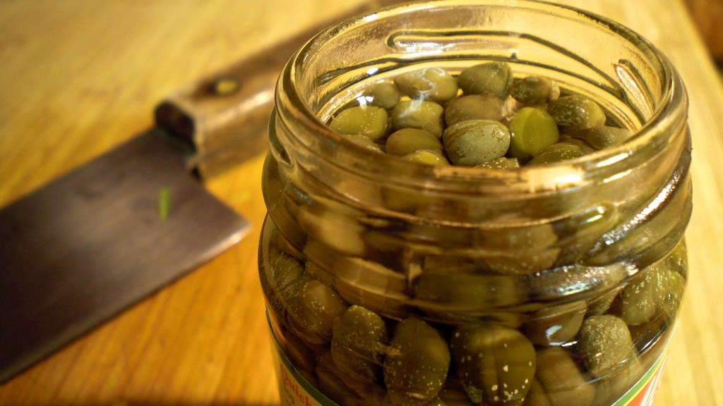
capers are the best food source of rutin
A recent headline proclaimed, "Mulberry extract activates brown fat, shows promise as obesity treatment." Beneath the headline, the text read:
Good news for those who want to activate their brown fat (or BAT, brown adipose tissue) without having to be cold: New research suggests that a natural compound in mulberries, called "rutin," can activate the BAT in our bodies to increase metabolism and facilitate weight loss.So I went to the research article on which the Science Daily article was based: "Rutin ameliorates obesity through brown fat activation."
Abstract
Increasing energy expenditure through activation of brown adipose tissue (BAT) is a critical approach to treating obesity and diabetes. In this study, rutin, a natural compound extracted from mulberry and a drug used as a capillary stabilizer clinically for many years without any side effects, regulated whole-body energy metabolism by enhancing BAT activity. Rutin treatment significantly reduced adiposity, increased energy expenditure, and improved glucose homeostasis in both genetically obese (Db/Db) and diet-induced obesity (DIO) mice. Rutin also induced brown-like adipocyte (beige) formation in subcutaneous adipose tissue in both obesity mouse models. Mechanistically, we found that rutin directly bound to and stabilized SIRT1, leading to hypoacetylation of peroxisome proliferator-activated receptor γ coactivator-1α protein, which stimulated Tfam transactivation and eventually augmented the number of mitochondria and UCP1 activity in BAT. These findings reveal that rutin is a novel small molecule that activates BAT and may provide a novel therapeutic approach to the treatment of metabolic disorders.We know the being cold can cause this activation of BAT, and we know that fish oil can also activate metabolism of brown fat. Rutin is the new kid on the block for this function.
I was intrigued, so I did a Google Scholar search for Rutin. Here are a few of the titles that came up:
- Rutin improves spatial memory in Alzheimer's disease transgenic mice by reducing Aβ oligomer level and attenuating oxidative stress and neuroinflammation
- Rutin prevents cognitive impairments by ameliorating oxidative stress and neuroinflammation in rat model of sporadic dementia of Alzheimer type
- Rutin inhibits UVB radiation-induced expression of COX-2 and iNOS in hairless mouse skin: p38 MAP kinase and JNK as potential targets
- Rutin and quercetin, bioactive compounds from tartary buckwheat, prevent liver inflammatory injury
- Rutin potentiates insulin receptor kinase to enhance insulin‐dependent glucose transporter 4 translocation
- Rutin decreases lipopolysaccharide-induced acute lung injury via inhibition of oxidative stress and the MAPK–NF-κB pathway
Rutin can act as a powerful antioxidant, especially with iron ions, preventing them from binding with hydrogen peroxide and preventing it from becoming a highly reactive free radical.
Furthermore, it has been shown to inhibit in vitro the vascular endothelial growth factor[9] in subtoxic concentrations, so acts as an inhibitor of angiogenesis. This finding may have potential relevance for the control of some cancers.Food sources of rutin - approximate rutin content per 100g of selected foods, based on the Phenol-Explorer database:
The cannabinoid mediated antidepressant activity of rutin shown in mice models employing weight-loaded forced swim test. Rutin treatment showed upregulation of CB1 receptors in mouse brain tissue demonstrating antifatigue activity and CB1 receptor-interacting proteins. Further, in brain tissues, an increase in expression of peroxisome proliferator-activated receptor-α coactivator (PGC-1α) and sirtuin 1 (SIRT1) was also demonstrated. Integrating together the cannabinoid, PPAR-γ, and opioid receptor activities, rutin may be a potential multitargeted polypharmacological agent in prevention and treatment of diseases involving dysregulation of PPAR and ECS.[10]
Animal research
While a body of evidence for the effects of rutin and quercetin is available in mice,[11] rats,[12] hamsters,[13] and rabbits,[14] as well as in vitro studies,[15] no clinical studies directly demonstrate significant, positive effects of rutin as dietary supplement in humans.
Hydroxyethylrutosides, synthetic hydroxyethyl acetylations of rutin, are used in the treatment of chronic venous insufficiency.
- Rutin inhibits platelet aggregation,[16] as well as decreases capillary permeability, making the blood thinner and improving circulation.[citation needed]
- Rutin shows anti-inflammatory activity in some animal and in vitro models.[17][18]
- Rutin inhibits aldose reductase activity.[19] Aldose reductase is an enzyme normally present in the eye and elsewhere in the body. It helps change glucose into the sugar alcohol sorbitol.
- Recent studies show rutin could help prevent blood clots, so could be used to treat patients at risk of heart attacks and strokes.[20]
- Some evidence also shows rutin can be used to treat hemorrhoids, varicosis, and microangiopathy.[21]
- Relatively high amount of rutin increases thyroid iodide uptake in rats and decreases serum T3 and T4 level. The decreased hormone level can be explained by its inhibitory effect produced on Thyroid peroxidase enzyme (TPO).[22]
- Rutin is also an antioxidant;[23] compared to quercetin, acacetin, morin, hispidulin, hesperidin, and naringin, it was found to be the strongest.[24][unreliable source?] [25] However, in other trials, the effects of rutin were lower or negligible compared to those of quercetin.[26][27]
- Rutin produces antinociceptive effects involving central modulation of the vlPAG descending circuit partly mediated by an opioidergic mechanism [28]
332 mg Capers, spiceA little more digging and I found this article, "The Pharmacological Potential of Rutin," by Aditya Ganeshpurkar and Ajay K. Saluja, in Saudi Pharmaceutical Journal, posted online 30 April 2016.
45 mg Olive [Black], raw
36 mg Buckwheat, whole grain flour
23 mg Asparagus, raw
19 mg Black raspberry, raw
11 mg Red raspberry, raw
9 mg Buckwheat, groats, thermally treated
6 mg Buckwheat, refined flour
6 mg Greencurrant
6 mg Plum, fresh
5 mg Blackcurrant, raw
4 mg Blackberry, raw
The following extract from the article documents the many and varied health benefits of rutin (with links to the original research articles). This is quite an impressive list.
2. Pharmacological actions
2.1. Central nervous system
2.1.1. Prevention of neuroinflammation
Rutin has demonstrated the neuroprotective effect on brain ischemia. Administration of rutin caused attenuation of ‘ischemic neural apoptosis’ due to the embarrassment of p53 expression and lipid peroxidation along with increment in ‘endogenous antioxidant defense enzymes’ (Khan et al., 2009). It has been found to be useful in hypoxic, glutamate and oxidative stress (Pu et al., 2007). Reduction of ‘neuroinflammation’ in rat model of ‘sporadic dementia of Alzheimer type’ (Javed et al., 2012) and neuroprotective effects in ‘dexamethasone-treated mice’ (Tongjaroenbuangam et al., 2011) were observed on rutin administration.
2.1.2. Promotion of neural crest cell survival
The neural crest is progenitor comprising of neural and mesenchymal potentials. Treatment of rutin to trunk neural crest cells increased their viability without altering cell differentiation and proliferation which could be due to modulation of ERK2 and PI3K pathways (Nones et al., 2012).
2.1.3. Sedative activity
CNS and behavioral activity of rutin were on hole board, thiopental-induced sleeping time and locomotor activity tests in mice. Rutin, given by intraperitoneal route caused a depressant action on the CNS. Research confirmed the CNS depressant activity of rutin was unlikely due to the involvement of GABAA receptor (Fernández et al., 2006).
2.1.4. Anticonvulsant activity
Rutin also possesses anticonvulsant activity and seems to be safe for patients with epilepsy as it does not alter the activity of any of the administered antiepileptic drugs nor demonstrates any adverse effects (Nieoczym et al., 2014).
2.1.5. Anti-Alzheimer activity and treatment of hyperkinetic movement disorder
Rutin suppressed activity of proinflammatory cytokines by diminishing TNF-α and IL-1β production in microglia. Such an effect seems to be useful in the treatment of Alzheimer’s disease as evident by prevention of β-amyloid oligomeric cytotoxicity (Wang et al., 2012). Rutin caused attenuation of streptozotocin-induced inflammation by decreasing the activity of the glial fibrillary acidic protein, interleukin-8, cyclooxygenase-2, inducible nitric oxide synthase and nuclear factor-kB and thereby prevented gross anatomical changes in rat hippocampus. Such an effect could be useful in averting cognitive deficits and proves to be beneficial in the treatment of ‘sporadic dementia of Alzheimer type’ (Javed et al., 2012).
‘Tardive dyskinesia’ is a motor disorder of the orofacial region raised due to chronic treatment with neuroleptic drugs, and is considered as a chief clinical concern in the treatment of schizophrenia. In a study, in haloperidol-induced orofacial dyskinesia, rutin treatment reversed behavioral changes such as orofacial dyskinetic movements, stereotypic rearing, locomotor activity, and percent retention along with restoration of biochemical and neurochemical parameters. Thus, rutin seems to be a lead molecule in the treatment of hyperkinetic movement disorder (Bishnoi et al., 2007).
2.1.6. Antidepressant effects
Forced swimming test and tail suspension test in mice were utilized to analyze ‘antidepressant-like effects’ of rutin isolated from Schinus molle. There was a reduction in the immobility time in the tail suspension test. There was no alteration in locomotor activity. Studies demonstrated the antidepressant-like effect of rutin mediated due to increasing the availability of serotonin and noradrenaline in the synaptic cleft ( Machado et al., 2008).
2.1.7. Stroke
Stroke could be regarded as a decisive public health issue that seems to be an important cause of fatality and disability in adults globally (Lloyd-Jones et al., 2009). Oxidative stress and inflammation are two of the pathological events observed after ‘ischemic injury’ in the brain (Deb et al., 2010). Protective effect of rutin an animal model of focal cortical ischemia induced by unilateral thermocoagulation of superficial blood vessels of the motor (M1) and somatosensory (S1) primary cortices was studied. Rutin administration significantly promoted recovery of sensorimotor loss which was observed due to the reduction of neurodegeneration in the periphery of cortical injury (Ortolani et al., 1995).
2.2. Analgesic and antiarthritic activities
2.2.1. Analgesic and antinociceptive effects
Analgesic effect of rutin was studied by hot plate test on swiss albino mice whereby the analgesic effect of rutin was established (Rylski et al., 1979). Further, it was also confirmed that rutin exhibited peripheral and central antinociceptive activities (Selvaraj et al., 2014).
2.2.2. Antiarthritic effects
Animals treated with rutin were observed with significant decrement in rheumatoid arthritis and Fanconi anemia by inhibiting ‘oxygen radical overproduction’ (Ostrakhovitch and Afanas’ev, 2001). In adjuvant arthritis rat model, rutin inhibited acute and chronic phases of inflammation. Rutin was the most active in the chronic stage of inflammation (Guardia et al., 2001). Due to antifungal and anti-arthritic effects, rutin has a therapeutic effect on septic arthritis caused by Candida albicans ( Han, 2009). Further in an independent study, rutin slowed down inflammatory and catabolic cartilage markers in osteoarthritic lesions in the Hartley guinea pig (Horcajada et al., 2014).
2.3. Endocrine system
2.3.1. Antidiabetic effects
Streptozotocin is a toxic chemical known to deplete levels of insulin by destroying pancreatic islets. Streptozotocin selectively assaults pancreatic β-cells by generating free radicals of oxygen and nitrogen monoxide along with reducing levels of NAD and NADP. Excessive production of glucose and its decreased utilization by tissues serve as the fundamental bases of hyperglycemia (Chattopadhyay, 1993). In a study, chronic administration of rutin in streptozotocin-induced diabetic rats caused a decrement in plasma glucose, augmentation in insulin levels, and restitution of glycogen content and glycolytic enzymes. Significant rejuvenation of pancreatic islets along with diminished fatty infiltrate was observed in rutin-treated diabetic rats (Stanley Mainzen Prince and Kamalakkannan, 2006 and Srinivasan et al., 2005). Diminution of fasting plasma glucose, glycosylated hemoglobin, C-peptide, and malondialdehyde levels was observed in rutin treated streptozotocin diabetic rats (Kamalakkannan and Prince, 2006). Rutin averted the levels of enzymes viz. ALT, AST, and LDH in the serum, liver, and heart demonstrating a protective effect on hepatic (Fernandes et al., 2010) and cardiac toxicity (Krishna et al., 2005) associated due to streptozotocin. Alteration in the activity of matrix metalloproteinase and protection to kidney against streptozotocin-induced damage was observed (Kamalakkannan and Stanely Mainzen Prince, 2006). Rutin stimulated glucose uptake in the soleus muscle, and effect was thought to be mediated through extracellular calcium and calcium-calmodulin-dependent protein kinase II activation. Increase in intracellular calcium concentration is involved in DNA activation which was mediated by rutin (Kappel et al., 2013). Rutin added for glycemic control via enhancement of insulin receptor kinase activity, thus aided in promoting ‘insulin signaling pathway’ that caused increased GLUT4 translocation and augmented glucose uptake (Hsu et al., 2014).
2.3.2. Anti-hypercholesterolemic effects
Rutin is a ‘selective and non-toxic modulator’ of hypercholesterolemia. In a study conducted on diet-induced hypercholesterolemic Golden Syrian hamster model, rutin significantly reduced plasma triglyceride levels in experimental animals (Kanashiro et al., 2009). Along with this, rutin also caused a decrement in levels of total cholesterol and HDL cholesterol (da Silva et al., 2001).
In a high-cholesterol diet fed male wistar rats, rutin administration demonstrated protective effect against the hepatotoxicity as evident by decrement of the plasma level of alanine transaminase (ALT), aspartate aminotransferase (AST), triglyceride (TG), total cholesterol (TC), and low-density lipoprotein (LDL) (Al-Rejaie et al., 2013). In another study, it was established that chronic consumption of flavonoids such as rutin could be favorable to cardiovascular health (Kalgaonkar et al., 2010).
2.3.3. Thyroid uptake promotion
‘Thyroid iodide uptake’ mediated via sodium-iodide symporter plays a pivotal role in thyroid hormone biosynthesis and also plays a key role in diagnosis and treatment of various thyroid diseases. However, some of the patients with thyroid cancer are obstinate to radioiodine therapy, which reduced iodine uptake ability, that remarkably reduces the probability of endurance. Thus, it becomes necessary to search for natural agents that aid in the uptake of thyroid iodide.
In a study, rutin caused a minor reduction in levels of serum T4 and T3 without changing serum thyrotropin. There was a significant increase in hypothalamic, pituitary and brown adipose tissue type 2 deiodinase along with the diminution of liver type 1 deiodinase activities. Administration of rutin was observed with increment in thyroid iodide uptake that could be due to an elevation in the activity of sodium-iodide symporter. The study demonstrates the effectiveness of rutin as an adjuvant in radioiodine therapy (Gonçalves et al., 2013).
2.4. Cardiovascular system
2.4.1. Hypertension
Buckwheat, a rich source of rutin is found to prevent oxidative damage in ‘aortic endothelial cells’ by lowering nitrotyrosine immunoreactivity. Germinated extract of buckwheat demonstrated antihypertensive effect and possibly shelter ‘arterial endothelial cells’ by detrimental effects of oxidative stress (Kim et al., 2009). Reduction in oxidative stress due to rutin, when administered by oral route, is the key reason for the restoration of ‘impaired baroreflex sensitivity’ and ‘vascular reactivity’ in hypertensive rats (Mendes-Junior et al., 2013). By augmenting NO production in human endothelial cells, rutin improved endothelial functions (Ugusman et al., 2014).
2.4.2. Blood coagulation
Chan et al. (2009) attempted to study effects of the rutin on the anticoagulant activity of oral warfarin and the protein binding along with pharmacokinetics of its enantiomers in rats. Rutin enhanced the in vitro serum protein binding of S- and R-warfarin. Rutin treatment significantly decreased the elimination half-life of S-warfarin by 37% as a result of the 69% increase in unbound clearance of the S-enantiomer. In a nutshell, concomitant administration of rutin possibly reduces the anticoagulant effect of racemic warfarin (Chan et al., 2009).
2.4.3. Antiplatelet aggregatory effect
Rutin in vitro caused concentration-dependent inhibition of platelet activating factor induced washed rabbit platelet aggregation, and intra-platelet free calcium concentration elevation was induced by platelet activating factor which was inhibited by rutin in a dose-dependent manner (Chen et al., 2002).
2.5. Gastrointestinal system
2.5.1. Antiulcer effects
A peptic ulcer is infirmity that influences the substantial population in the world. Ulcers are observed when disparity occurs among ‘aggressive’ and ‘protective’ factors at the luminal surface of the gastric epithelium. HCl, pepsins, nonsteroidal anti-inflammatory drugs, Helicobacter pylori, bile acids, ischemia, hypoxia, smoking, alcohol, etc. include dynamic features whereas defensive factors comprise of bicarbonate, a mucus layer, mucosal blood flow, PGs and growth factors ( Kalant et al., 2007).
Ethanol is ill-reputed agent known to produce damage to gastric mucosa in animal and clinical studies. Ethanol in a concentration higher than 400 ml/l causes significance in gross morphology of stomach which is observed by mucosal hyperemia, necrosis, edema and mucosal or submucosal hemorrhage (Oates and Hakkinen, 1988 and Szabo and Goldberg, 1990). Oxygen-derived free radicals could be regarded as a key reason for the formation of lesions (Pihan et al., 1987 and Szelenyi and Brune, 1988). Rutin pretreatment before administration of ethanol afforded significant protection against necrosis. Restoration in the levels of glutathione peroxidase along with ‘anti-lipoperoxidant effect’ was observed (La Casa et al., 2000). Similarly in an indomethacin-induced model of ulcers, rats pretreated with rutin caused reestablishment of altered oxidative stress and biochemical parameters possibly due to neutrophil infiltration, suppression of oxidative stress generation and replenishing nitrite/nitrate levels. Protective effects are also evident by histopathological investigations (Abdel-Raheem, 2010).
Another investigation provides insight into the molecular mechanism of action of rutin over gastric proton pumps. Rutin demonstrated concentration-dependent inhibition of goat gastric ATPase, with IC50 = 36 μg/ml, making it that rutin exerts an antiulcer effect by inhibiting the gastric proton pump (Dubey et al., 2013 and Dubey et al., 2013).
2.6. Respiratory system
2.6.1. Antiasthmatic activity and other associated effects
The antiasthmatic activity of rutin was studied in ovalbumin-sensitized conscious guinea pigs challenged with aerosolized ovalbumin where airway resistance during the immediate phase response and late-phase response was determined. Rutin significantly inhibited specific airway resistance and immediate-phase response along with reticence of histamine, phospholipase A2, and eosinophil peroxidase. There was reduced conscription of neutrophils and eosinophils into the lung (Jung et al., 2007). Use of rutin was also suggested in whooping cough along with vitamins C and K (De Sarmento and Kimura, 1957). In cats and whippets, rutin has been effectively used in the management of idiopathic chylothorax (Kopko, 2005, Gould, 2004 and Schuller et al., 2011).
2.7. Bones
2.7.1. Antiosteoporotic and antiosteopenic effect
Osteoporosis is a skeletal disorder observed by a decrement in bone strength, that is associated with augmented risk of fracture (Bhutani and Gupta, 2013). This condition is affecting elderly citizens globally and seems to be budding health predicament. Osteoporosis is observed in the case when bone resorption by osteoclasts surpasses bone formation by osteoblasts (Lau and Guo, 2011 and Roux, 2010). All therapeutic strategies for treatment of osteoporosis emphasize inhibition of ‘osteoclast-mediated bone resorption’. Parathyroid hormones are only classes of agents that ‘stimulate bone formation’ (Hodsman et al., 2005). In osteogenic-related assays, rutin caused proliferation and differentiation of human osteoblast-like MG-63 cells. There was also increase in the activity of alkaline phosphatase, expression of collagen type I and degree of mineralization (Hyun et al., 2014). Similar effects have been observed with rat calvarial osteoblast cells (Yang et al., 2006). Rutin inhibits osteoclast formation by decreasing oxygen reactive species and TNF-alpha by inhibiting activation of NF-kappaB (Kyung et al., 2008). Rutin inhibits ovariectomy-induced osteopenia in rats by slowing down resorption and increasing osteoblastic activity (Horcajada-Molteni et al., 2000). Thus, rutin can also be regarded as ‘osteoblast stimulant’.
2.8. Eye
2.8.1. Anticataract and ophthalmic effect
Formation of advanced glycation end products (AGE) is associated with cataract, a diabetic complication. Thus, inhibition of such glycation could be a useful strategy to prevent such complication. In this study, rutin caused embarrassment of glycation of proteins, chelation of metal complexes and partly inhibiting post-Amadori formation (Muthenna et al., 2010). In another study in wistar rat pups, selenium was used to induce cataract and protective effect of rutin in retarding cataractogenesis was investigated. Eye lens of rats treated with rutin were observed with restoration of lenticular antioxidant enzymes along with decrement in malondialdehyde formation. Experimental outcomes suggest that rutin prevented cataractogenesis possibly due to antioxidant mechanism (Isai et al., 2009).
Oral administration of rutin has been observed with an improved reduction in intraocular pressure (Vetrugno et al., 2012). Involvement of vitamins B1, B2, forskolin, and rutin demonstrates ‘defending effect’ on the ocular surface which aids to re-establish a ‘normal equilibrium’ of the tear film altered due to toxins (Nebbioso et al., 2013).
2.9. Excretory system
2.9.1. Diuretic effect
Quercetin, a metabolite of rutin, which is abundantly found in Hibiscus sabdariffa Linn acted on vascular endothelium causing nitric oxide release, leading to increasing renal vasorelaxation by increasing kidney filtration ( Alarcón-Alonso et al., 2012).
2.10. Reproductive system
2.10.1. Effect on sperm quality and male reproductive organs
Rutin, in a study, afforded protective effect on damage to human sperm induced by lipid peroxidation (Moretti et al., 2012). Rutin also demonstrated possible protection to testicular tissue and reproduction from oxidative stress observed in type 1 diabetes mellitus (Butchi Akondi et al., 2011) along with amelioration of cyclophosphamide-induced reproductive toxicity (Abarikwu et al., 2012) and testicular ischemia–reperfusion-induced oxidative stress in rats (Akondi et al., 2011).
2.11. Anticancer effects
Cancer includes a group of diseases that are characterized by the abnormal growth of cells that latently assault or spread to other parts of the body (Markert and Markert, 1968). Flavonoids are known to demonstrate an extensive assortment of biological effects, comprising of ‘antioxidant and radical-scavenging activities’. Reactive oxygen species have been associated with the pathogenesis of several diseases such as atherosclerosis and certain cancers.
Rutin has been extensively studied for anticancer/antineoplastic effects. In a study, human leukemia HL-60 cells were implanted in a murine model, and rutin (dose 120 mg/kg) caused a significant reduction in tumor size justifying antileukemic potential (Lin et al., 2012). In an independent study, rutin when administered to SW480 tumor cell lines (human colon cancer cell lines), was observed with less detrimental effects on the body and relative organ weight in mice along with an increment of mean survival time of 50 days (Alonso-Castro et al., 2013). Chem et al., demonstrated anti-neuroblastoma effect of rutin, where, rutin significantly inhibited the growth of LAN-5 cells and chemotactic ability. The study demonstrated that rutin could decrease BCL2 expression and BCL2/BAX ratio along with a reduction in levels of MYCN mRNA level and the secretion of TNF-α (C.Y. Chen et al., 2013). Rutin is also known to inhibit cancer cell growth by cell cycle arrest and/or apoptosis, along with inhibition of proliferation, angiogenesis, and/or metastasis in colorectal cell lines (Araújo et al., 2011). Rutin analog, quercetin, is studied against the proliferation of the ovarian cancer cell line OVCA 433, where dose-related inhibition was observed (Scambia et al., 1990). In another study, quercetin augmented apoptosis and barred metastasis in a model of pancreatic cancer (Mouria et al., 2002). Rutin also seems to be useful as an adjuvant in radioiodine therapy (Gonçalves et al., 2013).
2.12. Chemotherapeutic activity
2.12.1. Antibacterial activity
Rutin is extensively studied for antimicrobial activity against various strains of bacteria. It has demonstrated a profound degree of inhibition on growth of bacteria Escherichia coli ( Araruna et al., 2012). Rutin, quantified in honey has shown inhibitory effects over Proteus vulgaris, Shigella sonnei and Klebsiella sp. ( Pimentel et al., 2013). Antimicrobial activity against Pseudomonas auruginosssa and Bacillus subtilis has also been documented ( Dubey et al., 2013 and Dubey et al., 2013). In situ antimicrobial activity of rutin and other polyphenols in the food system has been studied, and the results demonstrate a promising involvement of flavonoids in the preservation of food ( Stojković et al., 2013).
Bernard et al., demonstrated that rutin by inhibiting DNA isomerase IV demonstrated antibacterial activity against E. coli ( Bernard et al., 1997). In a study, rutin synergistically enhanced antibacterial activity of other flavonoids against Bacillus cereus and Salmonella enteritidis. Minimum inhibitory concentration value for kaempferol was remarkably decreased by the addition of rutin ( Arima et al., 2002).
2.12.2. Antifungal activities
Rutin demonstrated antifungal activity against the strain of Candida gattii with a minimum inhibitory concentration of 60 μg/ml (Johann et al., 2011). It was suggested that chemical modification in rutin by the introduction of substitute group may alter physicochemical properties such as electron density, hydrophobicity and steric strain that may prove to be fruitful regarding increment of antifungal activity. Use of rutin in treatment of septic arthritis caused by C. albicans has also been suggested ( Han, 2009).
2.12.3. Antimycobacterial activity
In a study, flavonoids rich extract containing rutin demonstrated antimycobacterial activity against Mycobacterium smegmatis ( da Cruz et al., 2012).
2.12.4. Larvicidal activity
In a study, rutin significantly inhibited growth and propagation of larvae of S. aegypti where maximum mortality of larvae was observed at 72 h ( Dubey et al., 2013 and Dubey et al., 2013).
2.12.5. Antimalarial activity
Antimalarial effect of rutin in association with chloroquine was observed on white leghorn chickens infected with Plasmodium (Bennettinia) juxtanucleare ( Silveira et al., 2009). In another study, biochemical mechanism of the antimalarial activity of Azadirachta indica leaf extract revealed the presence of an abundant amount of quercetin, an active metabolite of rutin ( Iwu et al., 1986). Recently, the antiplasmodial activity of quercetin is documented against Plasmodium falciparum ( Ganesh et al., 2012).
2.12.6. Antiretroviral activity
In a study, sodium rutin sulfate, a sulfated rutin analog, was investigated for anti-HIV activity against HIV-1 X 4 viruses IIIB, HIV-1 R5 isolates Ada-M and Ba-L strains. The mechanism by which sodium rutin sulfate demonstrated antiretroviral effect was a blockade of viral entry and virus-cell fusion which was mediated via interaction with HIV-1 envelope glycoprotein (Tao et al., 2007).
2.12.7. Antiviral activity
Antiviral agents for the treatment of infections caused by retroviruses, orthomyxoviruses, herpes viruses, hepatitis B virus and hepatitis C virus are widely available (De Clercq and Field, 2006). Owing to the high incidence of viral infections along with no precise treatment for ‘appearance of new resistant viral strains’, its seems to be necessary to develop novel antiviral drugs.
Rutin has been tested against vesicular stomatitis virus on mouse fibroblasts and protected cells for about 24 h (Wacker and Eilmes, 1978). In the case of canine distemper virus infection, rutin affords immense viral embarrassment when added at the times of adsorption and penetration in the viral replicative cycle (Carvalho et al., 2013). Rutin a chief constituent of Capparis sinaica Veill demonstrated profound antiviral effect against avian influenza strain H5N1 using plaque inhibition assay in the Madin-Darby canine kidney ( Ibrahim et al., 2013).
2.13. Hair
Apoptosis of hair follicular cells is a leading cause of hair follicle degeneration. In a study, apoptosis of human follicular dermal papilla cells was observed after treatment with staurosporine; this event was completely introverted by exposure to rutin, spermidine, and zeaxanthin. There was the preservation of expression of anti-apoptotic molecules such as Bcl-2, MAP-kinases and their phosphorylated forms, which concludes the use of rutin in the prevention of apoptosis of hair follicular cells, one of the leading causes of baldness (Carelli et al., 2012).
2.14. Skin
2.14.1. Sunscreen effects
The desire for having healthy, beautiful and disease free skin is being observed from the eternal era. History of cosmetics and skin care product dates back in ancient Greek, Roman and Egyptian civilization. Advances in sciences and technology along with the development of modern chemistry have made this dream easy (Draelos, 2000). Exposure to ultraviolet B radiations produces ‘oxidative and inflammatory skin damage’ and augments jeopardy of skin ‘carcinogenesis. Rutin has been studied to explore the effects of rutin on UVB-induced inflammation in mouse skin in vivo. Topical application of rutin on mice skin 30 min before UVB irradiation reduced epidermal hyperplasia and the levels of proteins. There was also significant inhibition of UVB-induced expression of cyclooxygenase-2 (COX-2) and inducible nitric oxide synthase (iNOS) which could be due to inhibition of p38 MAP kinase and JNK that caused diminution UVB-induced expression of COX-2 in mouse skin (Choi et al., 2014). In a study conducted by Choquenet et al. (2008), rutin when incorporated in oil-in-water emulsions (concentration 10% w/w), sun protection factor (SPF) values comparable to ‘homosalate’ were observed. Along with TiO2, SPF values approached to 30.
2.14.2. In atopic dermatitis
In an atopic dermatitis model in BALB/c mice by repeated local exposure of house dust mite (Dermatophagoides farinae) extract (DFE) and 2,4-dinitrochlorobenzene (DNCB) to the ears, rutin treatment caused suppression of interleukin (IL)-4, IL-5, IL-13, IL-31, IL-32 and interferon (INF)-γ in the tissue. In 2,4-dinitroflourobenzene-sensitized a local lymph node assay for allergic contact dermatitis, rutin caused suppression of allergic contact dermatitis based on ear thickness and lymphocyte proliferation, serum IgG2a levels, and expression of INF-γ, IL-4, IL-5, IL-10, IL-17 and tumor necrosis factor-α in allergic contact dermatitis ears. This study signifies anti-atopic dermatitis and allergic contact dermatitis activity of rutin (Choi and Kim, 2013). Rutin has also demonstrated anti aging effects on the skin (Odetti et al., 1990). Thus, rutin could be regarded as most effective topically applied compound given its antioxidant activity and skin penetration profile (Alonso et al., 2014).
2.15. Immune effects
Vibrio alginolyticus challenge was studied based on immune and physiological response to rutin in white shrimp. In this study, physiological, innate non-specific immune responses, respiratory bursts and superoxide dismutase activity against the pathogen were analyzed. Rutin from Toona sinensis was administered to shrimp. In brief, shrimps treated with rutin maintained lower glucose, lactate, and lipid levels in response against the pathogen. Survival rates of shrimps treated with rutin were more ( Hsieh et al., 2008). Another study demonstrates promotion of the activity of macrophage phagocytosis in cells (Lin et al., 2009).
2.16. Body strength
2.16.1. Anti fatigue activity
Fatigue is an indication which is associated with person’s health. Fatigue is related to deterioration in ‘physical performance’. Extreme exercise is associated with accretion of surplus reactive free radicals that leads to an oxidative stress injury to the body. Depletion of energy along with the accumulation of excess metabolite is the key reasons for fatigue (Yu et al., 2010, Fitts, 1994, Coombes et al., 2002 and You et al., 2011). Rutin administration in mice prevented depletion of superoxide dismutase and reduced glutathione. After seven days of treatment with rutin, mice were sacrificed and analyzed for soleus muscle and brain for peroxisome proliferator-activated receptor-α coactivator and sirtuin one mRNA expression. Up-regulation of the CB1 cannabinoid receptor-interacting protein 1, myelin basic protein, Rho GDP dissociation inhibitor (GDI) alpha, and TPI indicated that rutin treatment inhibited condition anxiety via up-regulation of the expression of anxiety-associated proteins (Su et al., 2014) and hence in nutshell it can be regarded as treatment with rutin improves the various mutilations allied with physical fatigue.
2.17. Organ protective effects
2.17.1. Neuroprotective activity
In mice model, rutin inhibited oxaliplatin-induced chronic painful peripheral neuropathy. Oxaliplatin is one of the most important platinum compounds used in colorectal cancer chemotherapy. However, it suffers from a drawback of peripheral which seems to be difficult to treat. Rutin significantly decreased oxaliplatin-induced peroxidative changes in the spinal cord and lipid peroxidation along with inducible nitric oxide (Azevedo et al., 2013).
2.17.2. Retinoprotective activity
Effect of rutin on ocular blood flow by ‘colored microsphere technique’ was determined. Electroretinography was used to determine the b-wave recovery which is a tool for estimation of retinal function recovery. Rutin increased ocular blood flow and demonstrated a remarkable effect on retinal function recovery (Chiou and Xu, 2004).
2.17.3. Protective effect on lung tissue
Acute lung injury is severe disease observed with ‘high mortality and morbidity.’ Till date, there is no therapeutic stratagem established. Rutin demonstrated protective effects on histopathological changes in lung tissue along with prevention of ‘infiltration of polymorphonuclear granulocytes in bronchoalveolar lavage fluid’. There was also a reduction in secretion of lipid peroxidation and proinflammatory cytokines. The activity of antioxidant enzymes such as catalase, glutathione peroxidase, superoxide dismutase, and heme oxygenase-1 caused by lipopolysaccharide was reversed by rutin (Yeh et al., 2014). Pretreatment of rutin caused inhibition of lipopolysaccharide-induced arterial blood gas exchange and neutrophils infiltration in the lungs. There was suppression of macrophage inflammatory protein-2 and matrix metalloproteinase-9 (Chen et al., 2014). Another study demonstrated that rutin effectively inhibited vascular cell adhesion molecule-1 and inducible nitric oxide synthase (Huang et al., 2014). There is also evidence that rutin prevented early adult respiratory distress syndrome possibly due to inhibition of lipid peroxidation (Ortolani et al., 2000).
2.17.4. Cardioprotective effects
Ischemic heart disease is one of the primary reasons for ‘morbidity and mortality’ all along the globe. The pathophysiology underlying damage at a cellular level raised due to ischemia is multifaceted. ‘Reactive oxygen species’ are identified to participate and play a key task in the pathogenesis of several diseases (Li and Jackson, 2002). It is well known that ischemic tissues produce ‘oxygen-derived free radicals’ that are known to cause oxidative damage to membrane lipids, proteins, and carbohydrates that predispose to ‘qualitative and quantitative alterations’ of the myocardium (Burton et al., 1984). Rutin was studied for cardioprotective effect by ischemia–reperfusion-induced myocardial infarction in normal and streptozotocin diabetic rats. Rutin administration significantly offered cardioprotection and restricted infracts size in normal and diabetic rats (Annapurna et al., 2009 and Krishna et al., 2005). The mechanism lying behind this protection could be attenuation of lipid peroxidation in myocardial tissues (Ali et al., 2009).
Isoproterenol is β-adrenergic agonist used to treat bradycardia and heart block (Shen, 2008). However, frequent administration of it may lead to cardiac toxicity observed by an increase in the activity of serum cardiac marker enzymes viz lactate dehydrogenase, aspartate transaminase, creatine kinase, and alanine transaminase along with significant decrease in the activity of these enzymes in the heart. Rutin restored the levels of cardiac marker enzymes along with a reduction in lipid peroxidation. The study suggests the cardioprotective effect is due to the virtue of the antioxidant effect of rutin (Chan et al., 2009).
Repression of isoproterenol-induced increase in angiotensin II and aldosterone in plasma was observed after administered by rutin. Along with this, overexpression of transforming growth factor β1, connective tissue growth factor, and excessive deposition of extracellular matrix in isoproterenol-treated myocardial tissues was observed in the group of animals treated with rutin (Li et al., 2013).
2.17.5. Prevention of splenocyte apoptosis
Rutin is known to alter the viability and function of mitogen-stimulated splenocytes and thymocytes compared with non-stimulated cells. In a study, when splenocytes were cultured with mitogens, it was observed that there was a decrease in interferon-gamma production along with decrement in splenocyte apoptosis (Roseghini et al., 2007).
2.17.6. Hepatoprotective activity
Cirrhosis is the destruction of hepatocytes and following substitution with ‘scar tissue’ which alter the flow of blood through liver leading to the death of hepatocytes and n function of the liver (Wang and Nagrath, 2010). The event of liver damage is followed by ‘hepatic fibrosis’ whereby regeneration of apoptotic cells is observed after ‘repeated injury’ (Friedman et al., 2002). In the event, when the death of regenerating cells occurs, deposition of extracellular matrix is observed, which leads to deposition of ‘fibrillar collagen’ (O’Connell and Rushworth, 2008). Such pathology is difficult to treat. The effectiveness of treatment with colchicines, interferons, penicillamine and corticosteroids are contradictory and seem to be questionable. As oxidative stress is one of the causes beneath cirrhosis, use of antioxidants could be a unique strategy for treatment. Rutin is extensively studied for hepatoprotective activity in experimental animals. Khan et al., 2012a and Khan et al., 2012b evaluated the protective effect of rutin in carbon tetrachloride (CCl₄)-induced liver injuries in rats. Administration of rutin caused a decrement in levels of alanine aminotransferase, aspartate aminotransferase, alkaline phosphatase and gamma-glutamyl transpeptidase in serum raised due to carbon tetrachloride. The level of cholesterol in blood was regulated whereas the level of endogenous liver antioxidant enzymes such as catalase, superoxide dismutase, glutathione peroxidase, glutathione-S-transferase, glutathione reductase and glutathione was increased, whereas lipid peroxidation was decreased in a dose-dependent manner. Along with this, the activity of p53 and CYP2E1 was restored by treatment with rutin. In an independent study in BALB/c mice, rutin treatment allayed oxaliplatin-induced hepatotoxicity and neurotoxicity. Prevention of mechanical allodynia along with histopathological observations suggested the protective role of rutin in the prevention of hepatotoxicity (Schwingel et al., 2014).
2.17.7. Nephroprotective activity
Nephropathy is one of the most encountered pathogenesis observed in the populace all over the world regardless of age, caste, creed, race and environment. The reasons behind this condition result from anomalies rose due to different metabolic and physiological disturbances. Nephropathy is among the ten primary causes of death all along the globe (Zhang and Lindup, 1993). Gentamicin (Sonkar et al., 2014), ecstasy (Karami et al., 2014), cisplatin (Corcostegui et al., 1998), and carbon tetrachloride (Ogeturk et al., 2005) are some of the compounds known to demonstrate nephrotoxicity.
In a study, rutin demonstrated protective activity against oxonate-induced hyperuricemia and renal dysfunction in mice. Administration of rutin resulted in a the decrement in levels of serum urate, creatinine and blood urea nitrogen, serum and kidney uromodulin levels, and increased urine uromodulin, urate and creatinine excretion in hyperuricemic mice. There was a significant downregulation of mRNA and protein levels of mouse glucose transporter 9 and urate transporter 1, along with upregulation of mRNA and protein levels of organic anion transporter 1 and organic cation/carnitine transporters in the kidney of hyperuricemic mice. In a fructose-fed rat model for hyperuricemia, rutin blocked the NOD-like receptor three inflammasome activation and aided in the improvement in the signaling and reduced lipid accumulation in the kidney of rats (Hu et al., 2012) along with reversal of dysregulation of renal transporters (Hu et al., 2009). Inhibition of inducible nitric oxide synthase activity and reduction in 3-nitrotyrosine formation (Korkmaz and Kolankaya, 2013) along with inhibition of reactive oxygen species (Korkmaz and Kolankaya, 2010) in the kidneys seem to be an important approach to prevent renal ischemia/reperfusion injury.
In cisplatin-treated wistar rats, rutin pretreatment caused restoration of kidney function and oxidative stress biomarkers (Kamel et al., 2014). It partially inhibited effect on NFκB and TNF-α pathway mediated inflammation, caspase-3 mediated-tubular cell apoptosis, along with re-establishment of histopathological changes due to cisplatin administration (Arjumand et al., 2011). Rutin also demonstrated protective effects against potassium bromate-induced nephrotoxicity in rats. The decrement in DNA fragmentation, upregulation in the activity of antioxidant enzymes viz. catalase, superoxide dismutase, glutathione peroxidase, glutathione-S-transferase, glutathione reductase, and reduced glutathione along with a reduction in lipid peroxidation were observed (Khan et al., 2012a and Khan et al., 2012b).
2.17.8. Protective effect on blood vasculature
Rutin was found to resist damages in vascular beds due to a disturbance in myocardium supply leading to initial damage, however in a chronic model of ‘stenocardia in dogs,’ therapeutic effect of rutin is up to a limited extent (Pozin et al., 1996).
2.18. Protective effects on wounds
2.18.1. Wound healing activity
Rutin, formulated as hydrogel, when applied to skin lesions of rats, caused a decrease in wound area as compared to control hydrogels. There was a reduction in oxidative stress in wound area as signified by a reduction in lipid peroxidation and protein carbonyl content along with increased catalase activity (Almeida et al., 2012). Rutin-releasing chitosan hydrogels as injectable dressings for dermal wound healing have been formulated. These hydrogels promoted defined formation of neo-epithelium and thicker granulation, which is closer to the original epithelial tissue (Tran et al., 2011).
2.19. Radio modulatory effects
Radiation therapy is one of the principal strategies in anticancer therapy for treatment of various cancers; however, due to ill effects on normal tissues, its use is restricted. Ionizing radiations predispose to the generation of free radicals that damages DNA; cell death follows this event. Thus along with cancerous cells, the damage is observed with normal cells also. Some ‘radioprotectors’ have been screened; still, their clinical effectiveness is restricted due to toxicity with the frequent administration. In a study, rutin has been investigated for potential radioprotective effects. Rutin was administered to Swiss albino mice to assess its impact on ‘radiation-induced sickness along with the survival analyses’. Rutin at the dose of 10 mg/kg optimally demonstrated radioprotective effects. There was an increment in radiation tolerance and levels of antioxidant enzymes were restored along with a decrease in lipid peroxidation in liver (Patil et al., 2012). Similar protection by rutin against gamma radiations was also observed (Patil et al., 2013).


